Nine years ago, one of the world’s main artificial intelligence scientists highlighted an occupational species in danger of extinction.
“People should stop training radiologists now,” said Geoffrey Hinton, adding that it was “completely obvious” that in five years AI surpassed humans in that field.
Today, the radiologist, medical specialists in medical images that look inside the body to diagnose and treat diseases, still have a great demand. A recent study of the American College of Radiology projected a constant growth force until 2055.
Dr. Hinton, who received a Nobel Prize in Physics last year for pioneering research at AI, was generally right that technology would have a significant impact, but not as a work murderer.
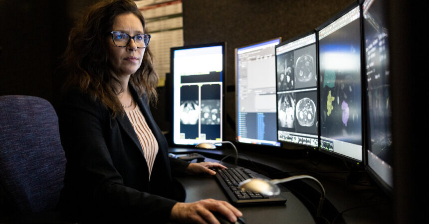
That is true for the radiologist at the Mayo Clinic, one of the main medical systems of the nations, whose main campus is in Rochester, Minnesota. There, in recent years, they have begun to use AI to sharpen images, automatic routine tasks, identify medical abnormalities and predict the disease. AI can also serve as “a second eye game.”
“But would radiologists replace? We did not believe it,” said Dr. Matthew Callstrom, president of Radiology of the Mayo Clinic, remembering the prediction of 2016. “We knew how difficult it is and everything that is involved.”
Informatics, labor experts and policy formulators have long discussed how AI will finally develop in the workforce. Will it be an intelligent assistant, improving human performance or a robotic substitute, displacing millions of workers?
The debate has intensified as avant -garde technology behind chatbots seems to be improving faster than expected. Openai, Anthrope and other companies in Silicon Valley now predict that AI will eclipse humans in most cognitive tasks in a few years. But many researchers prevent a more gradual in line transformation with the seismic inventions of the past, such as electricity or internet.
The planned radiologier extinction provides a case studies count. Until now, AI is verifying to be a powerful medical tool to increase efficiency and magnify human skills, instead of taking anyone’s job.
When it comes to developing and implementing AI in medicine, radiology has a main objective. Of the more than 1,000 AI applications approved by the Food and Medicines Administration for use in medicine, approximately three quarters are in radiology. The AI generally stands out to identify and measure a specific abnormality, such as a lung or breast lump.
“There has been surprising progress, but the thesis thesis tools mostly look for one thing,” said Dr. Charles E. Kahn Jr., Professor of Radiology at the Perelman Medicine Faculty of the University of Pennsylvania and editor of the magazine Radiology: Artificial Intelligence.
Radiologists do much more than studying images. They advise other doctors and surgeons, talk to patients, write reports and analyze medical records. After identifying a suspicious fabric group in an organ, they interpret what could mean for an individual patient with a particular medical history, taking advantage of years of experience.
The predictions that AI will steal often “will underestimate the complexity of the work that people do, as well as radiologists much more than reading scanns,” said David Author, Labor economist at the Massachusetts Institute of Technology.
In the May Clinic, artificial intelligence tools have been investigated, developed and adapted to adapt to the work routines of employed doctors. The staff has grown 55 percent from the forecast or fatality of Dr. Hinton, to more than 400 radiologists.
In 2016, stimulated by the warning and the advances in the recognition of images fed by AI, the leaders of the Department of Radiology gathered a group to evaluate the potential impact of technology.
“We think that the first thing we should do is use this technology to improve,” recalled Dr. Callstrom. “That was our first goal.”
They decided to invest. Today, the Radiology Department has a team of 40 people, including AI scientists, radiology researchers, data analysts and software engineers. They have developed a series of AI tools, from tissue analyzers to disease predictors.
That team works with specialists such as Dr. Theodora Potretzke, who focuses on the kidneys, bladder and reproductive organs. She describes the role of the radiologist as “a doctor for other doctors”, clearly communicating the results of the images, attending and advising.
Dr. Potretzke has collaborated in an AI tool that measures the volume of kidneys. Renal growth, when combined with cysts, can predict the decrease in renal function before appearing in blood tests. In the past, she measured the renal volume largely by hand, with the equivalent of a rule on the screen and conjectures. The results varied, and the task took a long time.
Dr. Potretzke served as a consultant, end user and tester while working with the AI team of the department. She helped design the software program, which has color coding for different fabrics and verified the measures.
Today, she mentions an image on the screen of her computer and clicks on an icon, and the measurement of renal volume appears instantly. He saves 15 to 30 minutes every time he examines a renal image, and is consistently precise.
“It is a good example of something that I feel very comfortable delivering AI to obtain efficiency and precision,” Dr. Ir. Potretzke said. “It can increase, help and quantify, but I am not in a place where I give up interpretive conclusions to technology.”
At the end of the hall, Dr. Francis Baffour, a staff radiologist, explained the varied ways in which the AI had applied the leg to the field, often in the background. Magnetic resonance scanners and CT manufacturers use AI algorithms to accelerate the images and to clean them, he said.
The AI can also automatically identify images that show the greatest probability of abnormal growth, they essentially tell the radiologist, “look here first.” Another program scan images for blood clots in the heart or lungs, even when the medical approach can be elsewhere.
“The AI is everywhere in our workflow now,” said Dr. Baffour.
In general, the Mayo Clinic is using more than 250 AI models, both internally developed and with suppliers license. The departments of Radiology and Cardiology are the greatest consumers.
In some cases, the new technology opens to ideas that are beyond human capacity. An AI model analyzes the data of electrocardiograms to predict patients most likely to develop atrial fibrillation, an abnormality of heart rate.
A radiology research project uses an AI algorithm to discern subtle changes in the form and texture of the pancreas to place cancer up to two years before conventional diagnoses. The Mayic team is working with other medical institutions to further try the algorithm in more data.
“Mathematics can see what the human eye cannot,” said Dr. John Halamka, president of the Mayo Clinic platform, which supervises the digital initiatives of the health system.
Dr. Halamka, an optimistic AI, believes that technology will transform medicine.
“Within five years, it will be Malpactic not to use AI,” he said. “But they will be human and the working together.”
Dr. Hinton agrees. In retrospect, he thinks he spoke too widely in 2016, he said in an email. Hey, it was not clear that I was talking exclusively about image analysis, and was at the time, but not in the direction, he added.
In a few years, most of the interpretation of medical images will be carried out with “a combination of AI and a radiologist, and will make the radiologists much more efficient in addition to improving accuracy,” said Dr. Hinton.